|
Pathology of Barrett's Esophagus
1. Multilayered Epithelium:
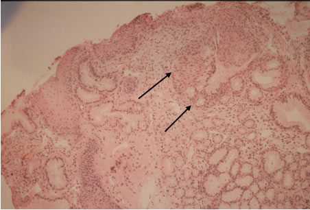
100X
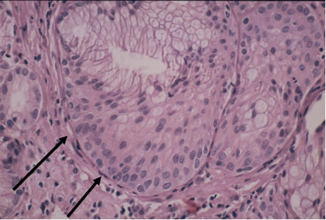
400X
Multilayered epithelium is composed
of squamoid cells at the base and columnar cells at the top. It has been
noted in Barrett’s
esophagus and carditis related to GERD, but not H. pylori infection. It may
represent a transitional
stage in the development of Barrett's esophagus. See references:
Odze, RD, et al., Surgical Pathology of the GI Tract, Liver, Biliary Tract
and Pancreas (Philadelphia, PA: Saunders, 2004)
Shields HM, Rosenberg SJ et al., “Prospective evaluation of multilayered
epithelium in Barrett's esophagus,” Am J Gastroenterol. 2001 Dec; 96(12):3268-73
Patient A: 70 year old female with a long history of heartburn. Endoscopy
showed a large Hiatal hernia and a 7 cm segment columnar mucosa.
2. Barrett’s esophagus
(low power):
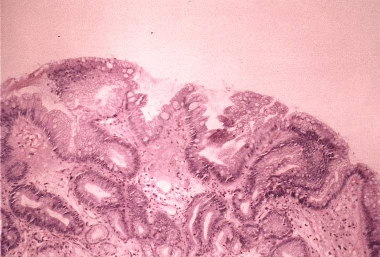
Figure 1
Slightly villiform glandular mucosa with intestinal metaplasia and antral
type gastric glands has replaced the normal squamous mucosa of the esophagus.
The intestinal metaplasia is established by the presence of the goblet
cells with the distinctly ovoid mucin droplets, in comparison to the smaller mucin
vacuoles of the foveolar mucinous cells. This type of intestinal metaplasia
is considered incomplete type. In complete type of intestinal metaplasia, goblets
cells, as well as small intestinal absorptive cells characterize the epithelium.
3. Barrett’s esophagus (high power):
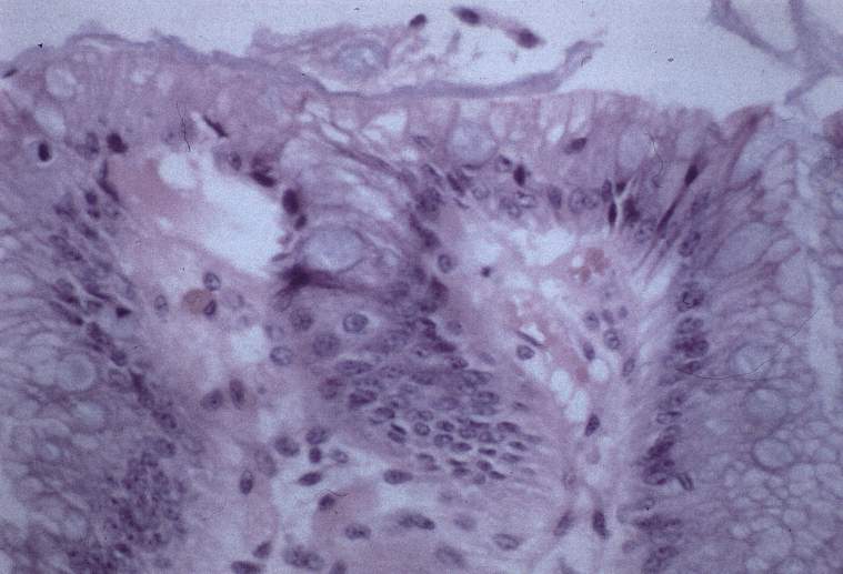
Figure 2
Incomplete intestinal metaplasia: goblet cells and foveolar gastric type epithelium
cells.
4. Barrett’s esophagus (high power):
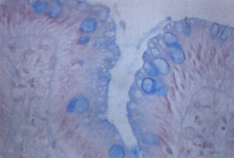
Figure 3
Goblet cells are also identified by their content of acid mucins, made visible
by the blue staining reaction with alcian blue at pH 2.5.
Pathology slides of our patients were provided by Dr. James P. Kolton of Caritas
Norwood Hospital, Norwood, MA.
|